Eye Surgery · Medical Tourism in Mexico
Your vision and your trip,
planned with the same precision.
From your first online consultation to your recovery, we coordinate every detail of your eye surgery in Mexico — so you can arrive with confidence and focus only on seeing better.
Subspecialty — Anterior Segment Microsurgery
Affiliated with — Asociación para Evitar la Ceguera en México IAP
Initial evaluation — 100% online, no travel needed yet
The process
Four steps, one well-planned trip.
This is how we guide every patient who travels to Mexico for eye surgery, from the first video call to final discharge.
01
Online consultation
We review your case and visual history, and answer your questions from wherever you are.
02
Travel plan
We help you plan your dates, lodging, and transportation ahead of time.
03
Surgery
Your procedure is performed following microsurgery protocols, by specialists.
04
Recovery and follow-up
We closely follow up on your recovery before you head back home.
Services
Complete eye care, for adults and children.
From diagnosis to surgery and optical care, with specialized attention for infants, children, and adults, all under one team in Bucerías, Nayarit.

Eye care for adults and children
General assessment and diagnosis, with state-of-the-art equipment for the whole family.
See details →
Cataract surgery
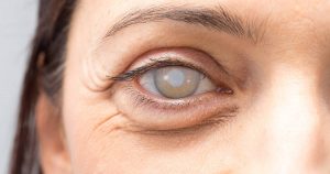
Lens replacement with precise calculation of the intraocular lens to be implanted.
See details →
Glaucoma: diagnosis and treatment
Early detection, monitoring, and treatment with drops, laser, or surgery, depending on each case.
See details →
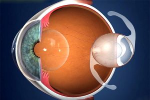
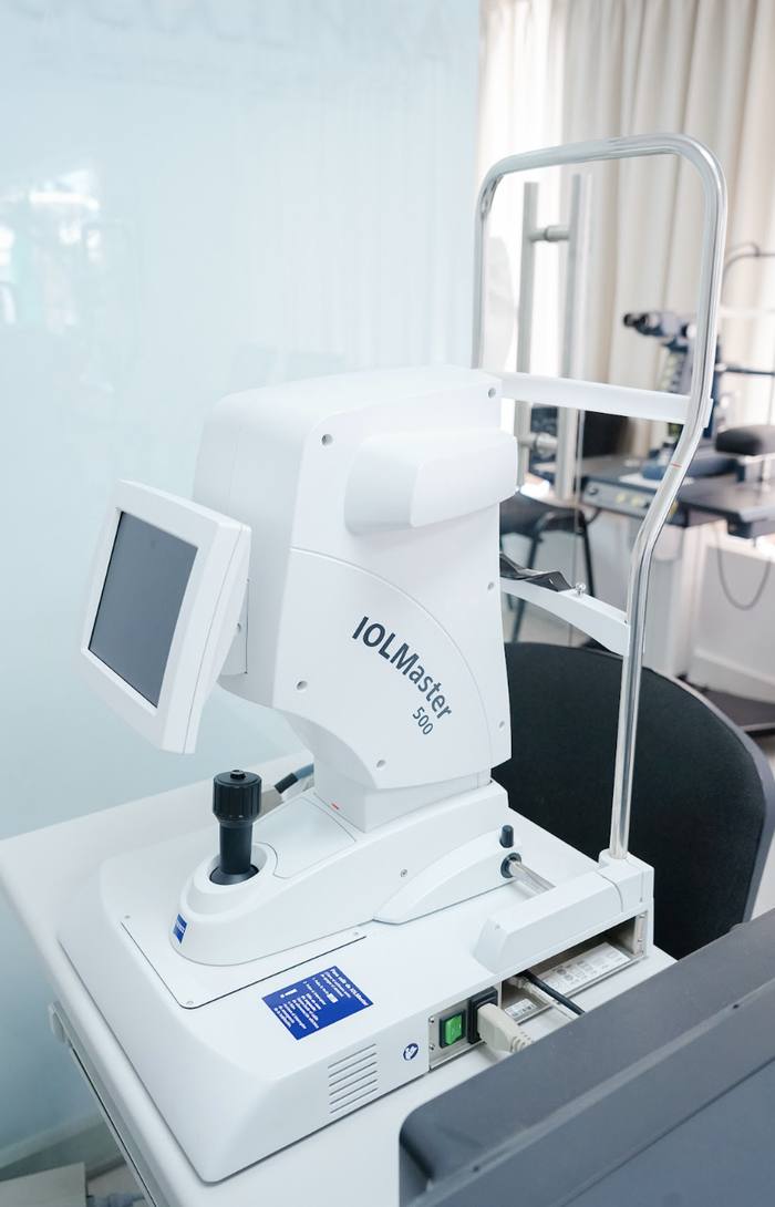
Intraocular lens power calculation
Non-invasive biometry, an essential part of pre-op assessment for cataract surgery.
See details →
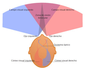
Visual field test
Assessment of peripheral vision through confrontation testing or tangent screen exam.
See details →
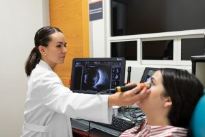
Ocular ultrasound
Imaging to assess internal eye structures when direct visualization isn't enough.
See details →

YAG laser
Outpatient laser procedure for capsulotomy, iridotomy, and other precise treatments without an incision.
See details →

Pediatric ophthalmology
Early detection and treatment of vision problems in infants and children, for healthy visual development.
See details →

Optical shop
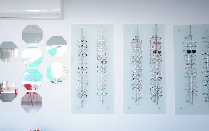
Prescription, contact, sun, polarized, and Transitions lenses — single and bifocal, for the whole family.
See details →
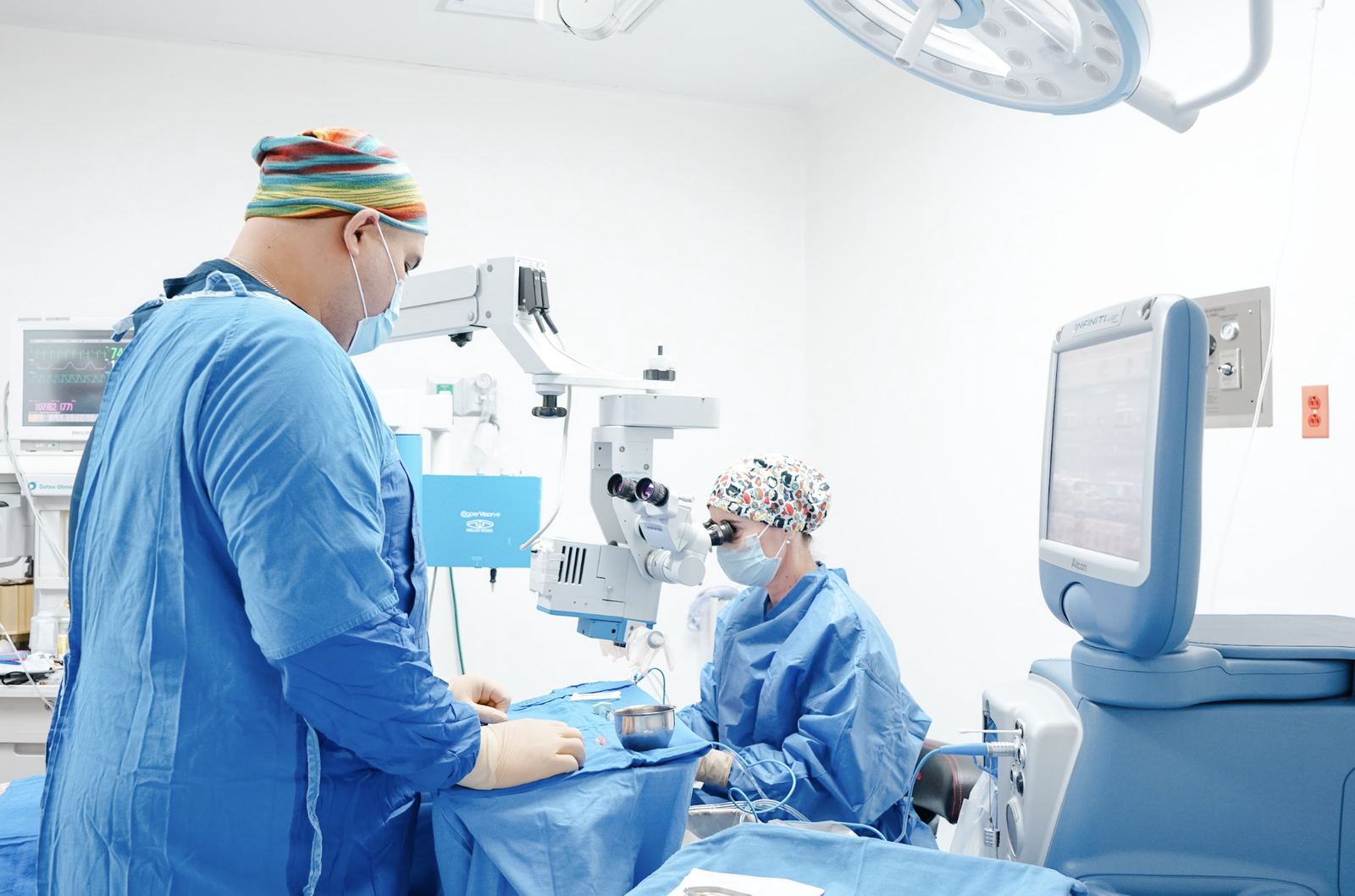
Our facilities
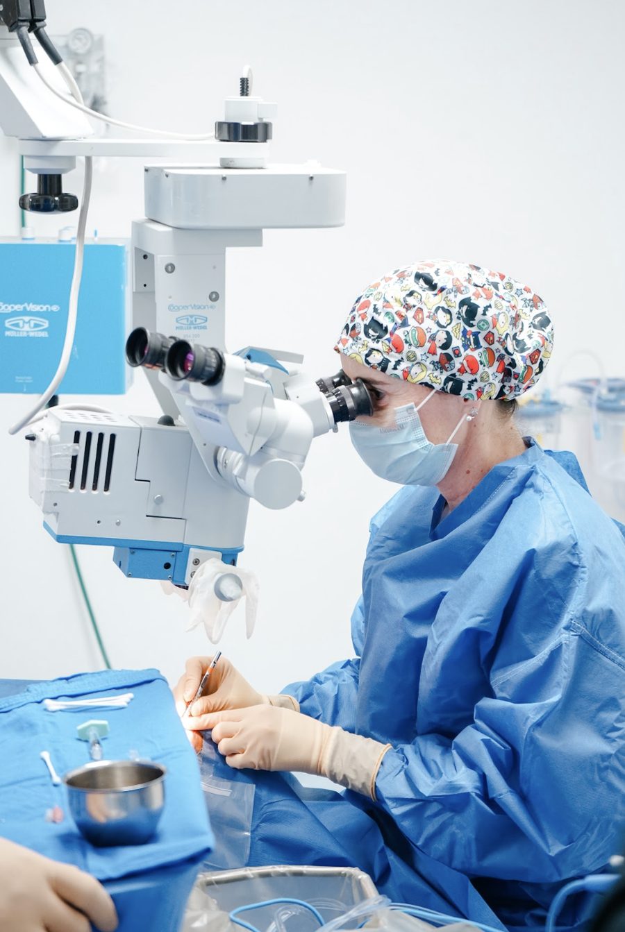
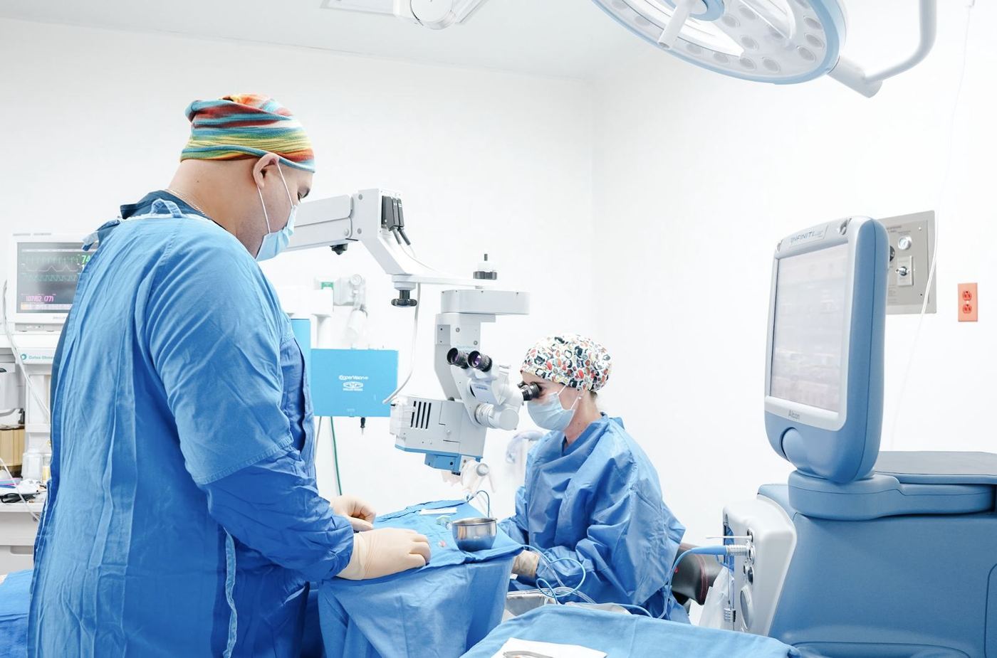
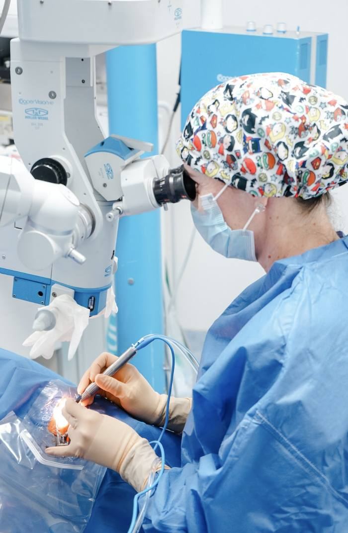
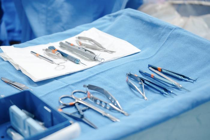
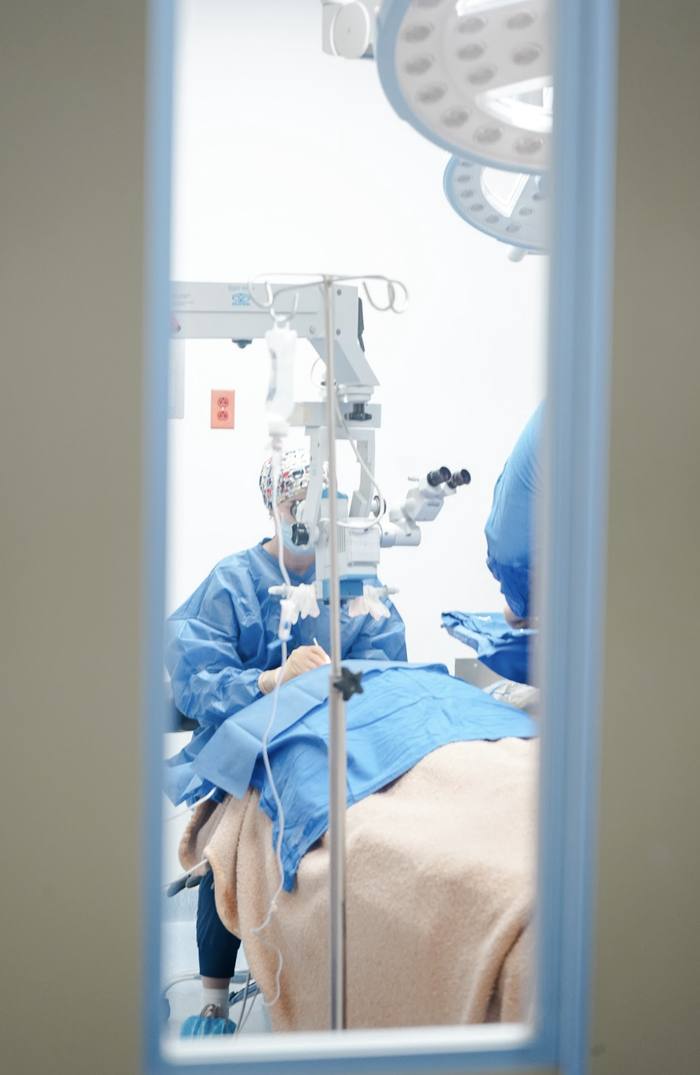
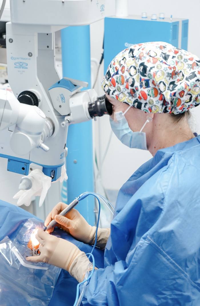
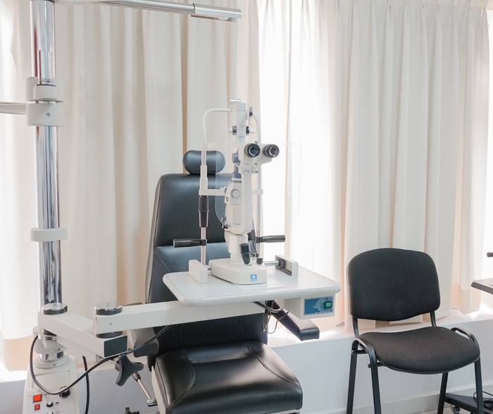
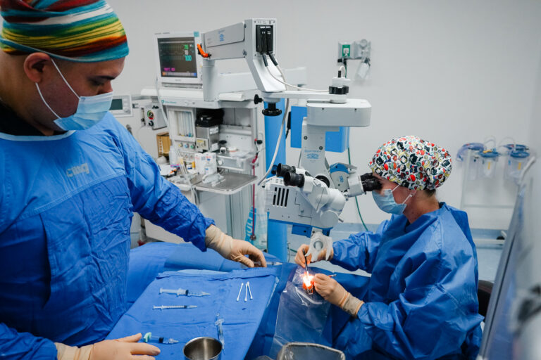
An operating room equipped with cutting-edge technology.
Gallery
A look inside our facilities.
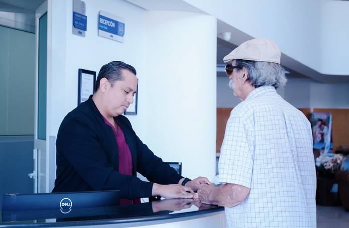
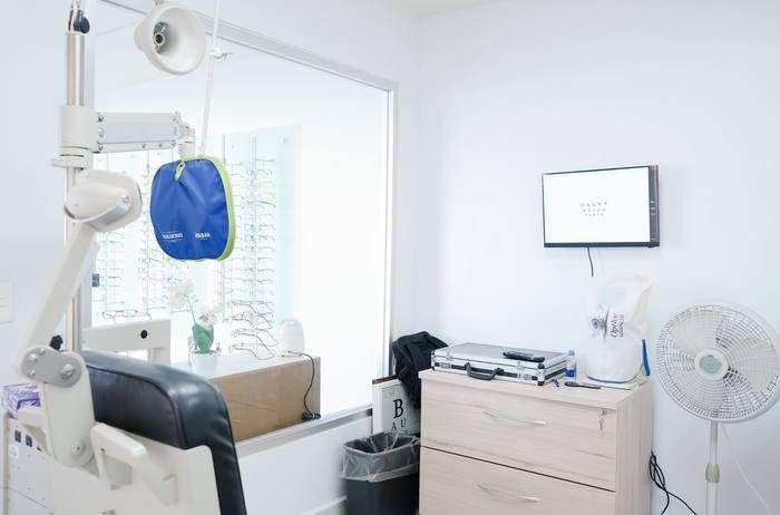
From the reception area to the operating room, this is Ocuclinika from the inside.
Why Ocuclinika
More than surgery: a trip handled from start to finish.
Full coordination
We organize your consultation, surgery, and travel logistics as a single process.
Focused subspecialty
Anterior Segment Microsurgery, affiliated with the Asociación para Evitar la Ceguera en México IAP.
Personal attention
Personalized follow-up before, during, and after your procedure.
Transparency
Clear information about your diagnosis, options, and costs, with no surprises.
“Let choosing what to pack be the hardest part of the trip.”
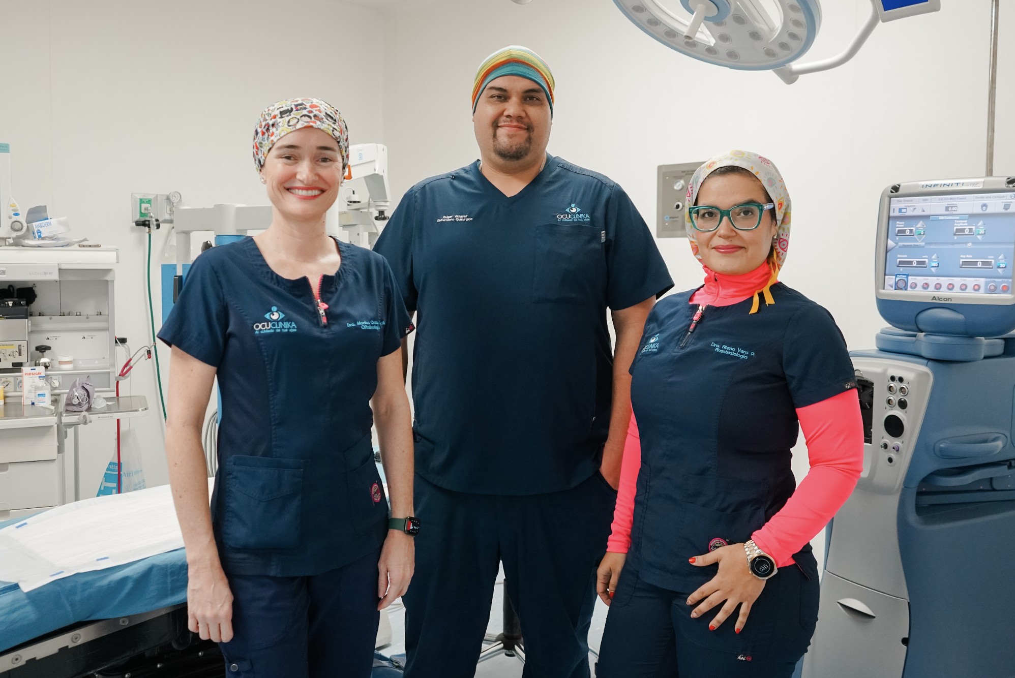
Medical team
The specialists behind your eye care.
A certified team, trained at some of Mexico's most respected ophthalmology institutions.
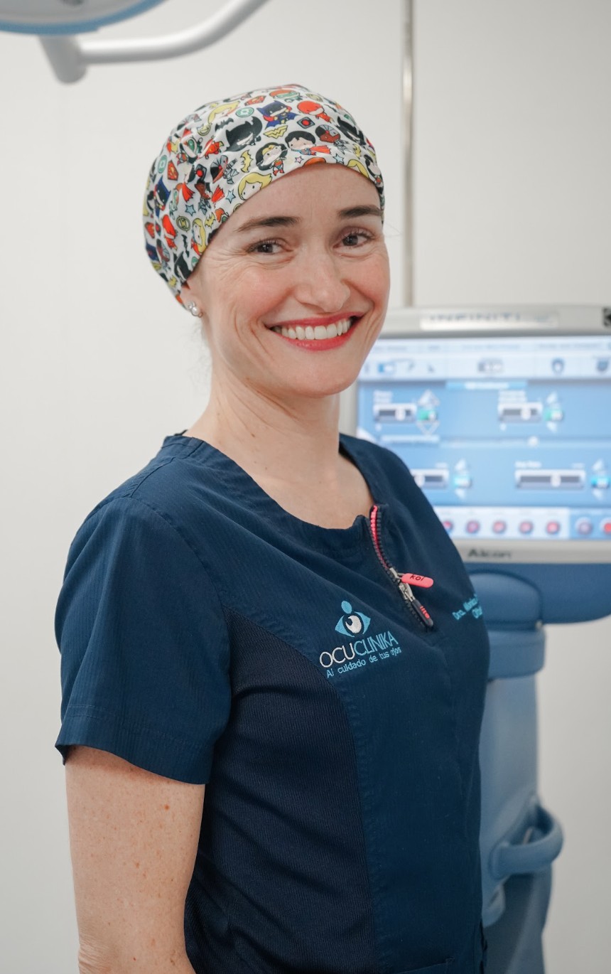
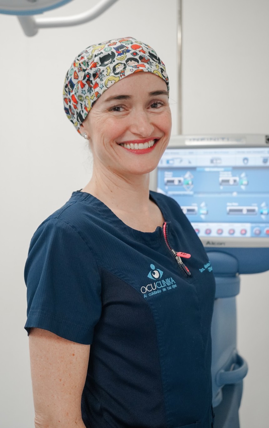
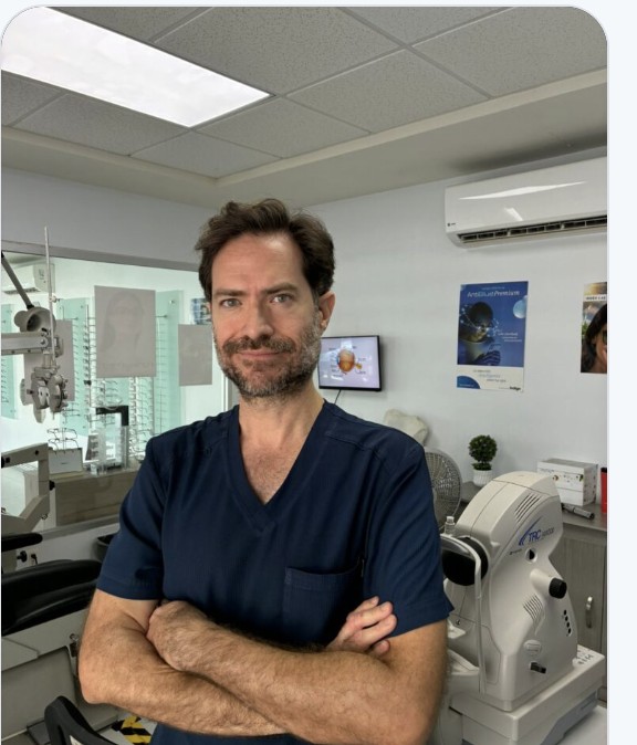
Dr. Mónica Orozco Carroll
Ophthalmology
Ophthalmologist with over 15 years of experience, specialized in Anterior Segment Microsurgery. Leads clinical care at Ocuclinika, caring for the eye health of patients of all ages.
- Medical Doctor — Universidad Autónoma de Guadalajara (UAG)
- Specialty in Ophthalmology — APEC, "Dr. Luis Sánchez Bulnes" Hospital
- Subspecialty in Anterior Segment Microsurgery — APEC
- Ophthalmology and Embryology Professor — UNIVA
- Member of ASCRS · "Dr. Luis Sánchez Bulnes" Award, 2008
🎙️ Listen to her on the radio
Pending: add the real radio interview link here
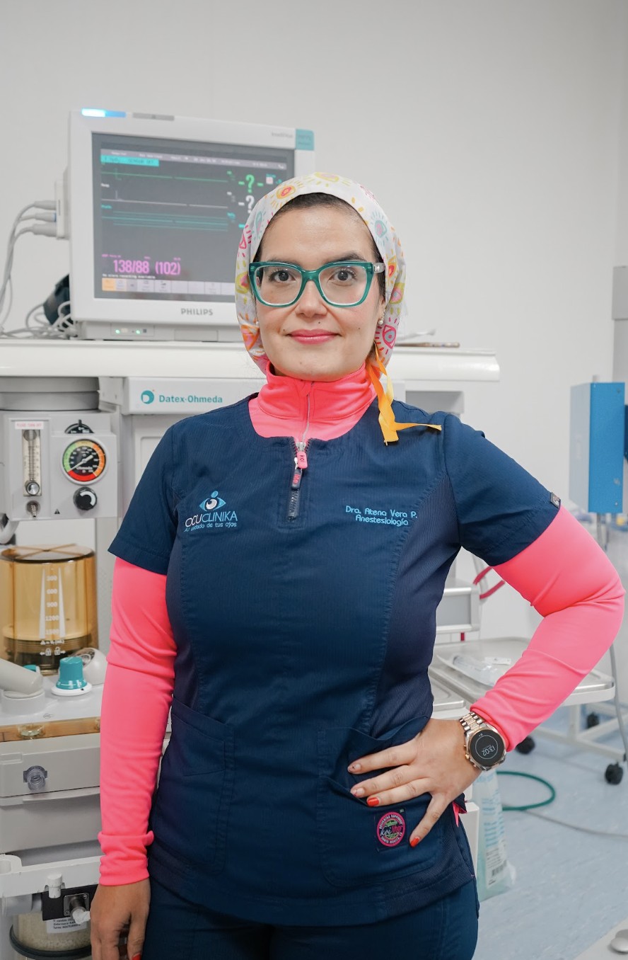
Dr. Atena Leonor Vera Ponce
Anesthesiology
Responsible for anesthesia in Ocuclinika's surgical procedures, in addition to her teaching work in ophthalmology.
- Medical Doctor — Universidad Autónoma de Guadalajara (UAG)
- Specialty in Anesthesiology — Universidad Autónoma de Chihuahua
- Ophthalmology Professor — UNIVA, since February 2016
Dr. Gilberto Moreno Ahumada
Ophthalmology · Strabismus
Specialist in the treatment of eye misalignment (strabismus) in patients of all ages, in the detection of vision problems in children and adults, and in the rehabilitation of patients with amblyopia. Also specialized in the surgical management of cataracts.
- Medical Doctor — Universidad de Guadalajara
- Advanced specialty in Strabismus — APEC, 2010
- Advanced specialty in Anterior Segment Microsurgery — Instituto Nacional de Rehabilitación (INR), 2011
Location
Visit us in Bucerías, Nayarit.
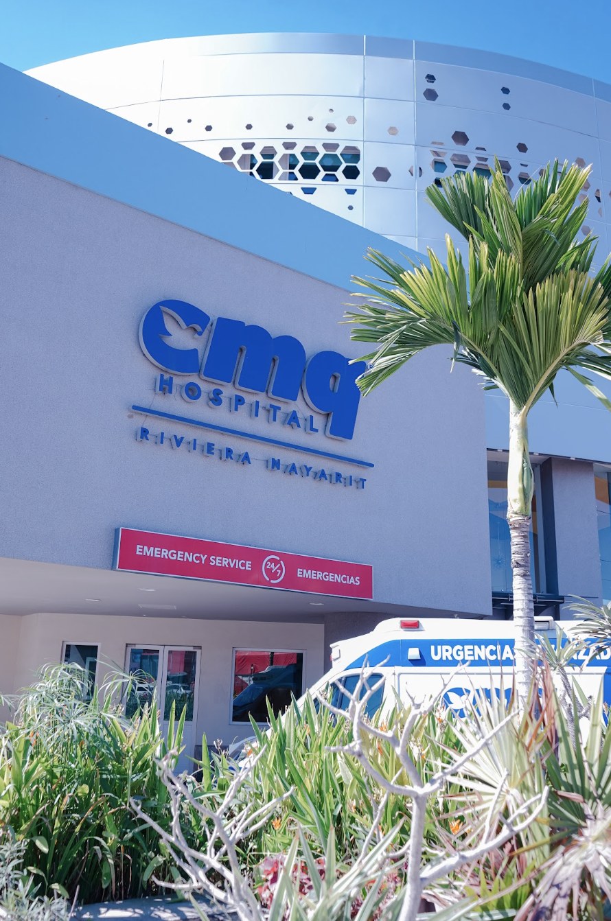
Inside CMQ Hospital Riviera Nayarit, minutes from Puerto Vallarta — easy to find and coordinate if you're traveling from outside Mexico.
General ophthalmic assessment and diagnosis for the whole family, using state-of-the-art equipment. We evaluate visual acuity, eye health, and any symptom that concerns you, for adults and children of any age.
Replacement of the clouded natural lens with an intraocular lens, with prior calculation of its optical power to aim for the best possible visual outcome. A routine procedure within our anterior segment microsurgery practice.
Glaucoma is a disease that can slowly damage the optic nerve, which carries information from the eye to the brain. This damage is usually related to elevated pressure inside the eye, although it can also occur when pressure is within normal values.
What matters most is that it rarely causes symptoms at first. A person may seem to see well and not realize they are losing vision. Over time, glaucoma tends to take away side vision first and, if left untreated, it can cause significant vision loss.
The good news is that glaucoma can be controlled. With drops, laser, or surgery, depending on each case, we can reduce the risk of further damage to your vision. That is why regular eye exams matter so much, especially if you have relatives with glaucoma, are over 40, or have other risk factors.
Treatment does not aim to restore vision that has already been lost, but to protect the vision you still have.
What symptoms does it cause?
The most important thing to know is that glaucoma often causes no symptoms at all in the beginning. That is why a person can have glaucoma and feel that they see perfectly well.
As the disease advances, side vision may gradually be lost, but the patient may not notice because the brain adapts to compensate for that loss. In more advanced stages, vision may feel more limited, or it may become hard to see objects off to the sides.
This is why we should not wait for symptoms to detect glaucoma. Regular exams with your ophthalmologist are essential to find it in time and protect your vision.
When to seek emergency care
There is a type of glaucoma that can appear suddenly. In those cases you may experience intense eye pain, a red eye, blurred vision, rings or halos around lights, headache, nausea, or vomiting. If these symptoms come on suddenly, seek emergency ophthalmic care.
How is it diagnosed?
There is no single test that on its own confirms or rules out glaucoma. Your ophthalmologist performs several studies and brings the results together.
- Eye pressure measurement. A quick test to find out the pressure inside the eye. Depending on the equipment, a device gently touches the surface of the eye or uses a small puff of air. It is generally painless and takes only a few seconds.
- Optic nerve examination. The ophthalmologist examines the back of the eye with different instruments. Drops are sometimes used to widen the pupil for a better view. This shows whether the nerve that carries information from the eye to the brain has any change related to glaucoma.
- Visual field test. This checks how well you see to the sides. You sit in front of a device and are asked to look steadily at a central point while small lights appear in different places; you press a button each time you see one. It is important to keep looking at the central point and respond only when you actually see a light.
- OCT of the optic nerve and retina. Like taking a highly detailed photograph of the inner layers of the eye. You rest your head against the device and look at a point of light; the equipment captures the images without touching the eye and obtains very precise information in seconds.
- Gonioscopy. This lets us examine the area where fluid drains out of the eye. After anesthetic drops are applied, the ophthalmologist gently places a small lens on the surface of the eye. You may feel slight pressure, but it should not be painful.
Why several studies? Each test gives us different information. Putting the results together tells us whether glaucoma is present, whether there is optic nerve damage, and whether the disease is stable or has changed over time. Not every patient needs exactly the same studies: the ophthalmologist selects them according to each person's characteristics.
How is it treated?
Glaucoma has no cure that restores lost vision, but treatments exist that can stop or slow the progression of the disease. The main goal is to lower the pressure inside the eye to a level that is appropriate for each patient.
- Eye drops. The most widely used treatment. One or more drops a day help lower eye pressure. It is very important to use them every day exactly as prescribed, even when you feel well, because glaucoma can progress without causing any discomfort.
- Laser. In some patients we use a laser to help fluid drain from the eye more easily and lower the pressure. It is usually performed in the office, takes a few minutes, and does not require conventional surgery.
- Surgery. When drops or laser are not enough, surgery may be necessary. Some procedures create a new pathway for fluid to leave the eye; others use small devices to help it drain. There are also minimally invasive glaucoma surgeries, suitable for certain patients.
Which treatment do I need? Not every patient needs the same one. The choice depends on the type of glaucoma, how advanced it is, the eye pressure, the condition of the optic nerve, and other individual factors.
The key point is that treatment is meant to protect the vision we still have. That is why, even when a patient sees well and has no discomfort, they should continue with their check-ups and treatment. Glaucoma is a disease we can control, but it requires lifelong follow-up.
Intraocular lens (IOL) calculation with the IOLMaster is an optical biometry method that obtains the measurements needed to calculate the power of the lens to be implanted during cataract surgery.
What to expect during the test
This is a non-invasive study: nothing touches the patient's eyes, it is painless, and it takes about five minutes.
What does the IOLMaster measure?
The device mainly obtains:
- Axial length (AL): the distance from the cornea to the retina.
- Keratometry (K1 and K2): the curvature of the cornea along its principal meridians.
- Anterior chamber depth (ACD): the distance from the cornea to the natural lens.
- Depending on the model, it may also measure white-to-white (WTW), pupil diameter, and other biometric parameters.
How is the calculation performed?
- Biometric measurement. The patient fixates on the IOLMaster target while the device takes several measurements.
- Quality check. The repeatability of the measurements is verified, along with the reliability of the axial length and keratometry readings.
- Formula selection. The data are entered into an IOL calculation formula. Modern formulas include, among others, Barrett Universal II, Kane, EVO, Haigis, and SRK/T.
- Refractive target selection. The desired refraction after surgery is set — for example emmetropia, residual myopia, or monovision.
- IOL power calculation. The formula uses axial length, corneal power, and parameters related to the effective lens position (ELP) to determine the IOL power.
- Comparison of results. It is advisable to compare several formulas, especially in eyes with extreme axial length, previous corneal refractive surgery, atypical corneas, or situations that make biometry difficult.
Clinical importance
The accuracy of the calculation depends fundamentally on the quality of the biometry, particularly the axial length and keratometry. A small error in axial length can produce a significant postoperative refractive error.
In patients with a dense cataract, when optical biometry cannot obtain a reliable measurement, ultrasound biometry may be required.
In short: the IOLMaster measures the anatomical characteristics of the eye and, through a biometric formula, calculates the IOL power needed to reach the refractive target chosen before cataract surgery.
Automated visual field testing is an ophthalmic study that measures the visual field: that is, how well a person perceives light stimuli appearing in different areas while keeping their gaze fixed on a central point.
How is it performed?
The patient sits in front of a device called a perimeter, generally with one eye tested at a time. They must keep their gaze fixed on a central point while small light stimuli appear in different parts of a screen or inside a dome.
Each time the patient perceives a stimulus, they respond by pressing a button. The equipment automatically varies the intensity and location of the lights to determine the sensitivity threshold at different points of the visual field.
What information does it provide?
The result makes it possible to identify:
- Visual field defects or losses.
- Their location and extent.
- The sensitivity of different areas of the retina and the visual pathway.
- Changes across successive studies, to determine whether there is progression.
It is especially important in diagnosing and monitoring glaucoma, but it can also be used in diseases of the retina, optic nerve, and visual pathway, as well as in some neurological conditions.
What does the result mean?
The study produces a sensitivity map of the visual field, along with various indices and graphs. Areas of lower sensitivity may appear as zones of depression or as scotomas.
The result must be interpreted together with the eye examination, intraocular pressure, optic nerve appearance, and structural studies such as OCT, since an abnormal visual field does not by itself necessarily mean that glaucoma is present.
It is also a psychophysical test: it depends on the patient's attention, understanding, and cooperation. Factors such as fatigue, poor fixation, refractive errors, or inconsistent responses can therefore alter the result.
B-scan ocular ultrasound is an imaging study that uses high-frequency ultrasound waves to obtain a two-dimensional image of the internal structures of the eye and the orbit. It is especially useful when the ocular media do not allow the posterior segment to be adequately visualized through ophthalmoscopy.
What to expect during the test
This is a non-invasive, painless study. The transducer is placed on the patient's eyelids with the eyes closed, and it takes between five and ten minutes.
What is it used for?
It mainly allows us to evaluate:
- Vitreous and retina: identifying retinal detachment, vitreous hemorrhage, and posterior vitreous detachment.
- Choroid: including choroidal detachments and some tumoral lesions.
- Natural lens: when significant opacity is present.
- Optic nerve and orbital structures in certain indications.
- Intraocular or orbital masses, foreign bodies, and other structural abnormalities.
Main indications
It is particularly important in patients with advanced cataract, vitreous hemorrhage, corneal opacity, or any other condition that prevents examination of the fundus. It is also used to assess ocular trauma and suspected retinal detachment.
In short: B-scan provides a dynamic, two-dimensional image of the globe and the orbit, making it possible to assess structures that cannot be observed directly and to detect posterior segment abnormalities.
YAG (Yttrium-Aluminum-Garnet) laser is an ophthalmic laser that uses high-energy light pulses to create small, precise openings or cuts in eye tissue, without the need for a conventional surgical incision.
Main indications
- YAG capsulotomy: treatment of posterior capsule opacification after cataract surgery. This is its most common indication and it restores clarity to the visual axis.
- YAG peripheral iridotomy: creates a small opening in the iris to facilitate the flow of aqueous humor, mainly indicated for angle-closure glaucoma or patients at risk of angle closure.
- Hyaloidotomy or membranotomy: in select cases of premacular or subhyaloid hemorrhage, to allow blood to drain into the vitreous cavity.
It's an outpatient procedure, quick and generally painless, though it requires a prior eye exam and follow-up due to possible complications such as increased intraocular pressure, inflammation, or, rarely, retinal changes.
Early detection and treatment of vision problems in infants and children, to support healthy visual development from an early age.
Prescription, contact, sun, polarized, and Transitions lenses, with single-vision and bifocal options for the whole family. We'll help you choose the frame and lens type that best fits your needs.